MINIMALLY INVASIVE AESTETHIC REHABILITATION OF THE ANTERIOR SECTORS WITH PORCELAIN VENEERS
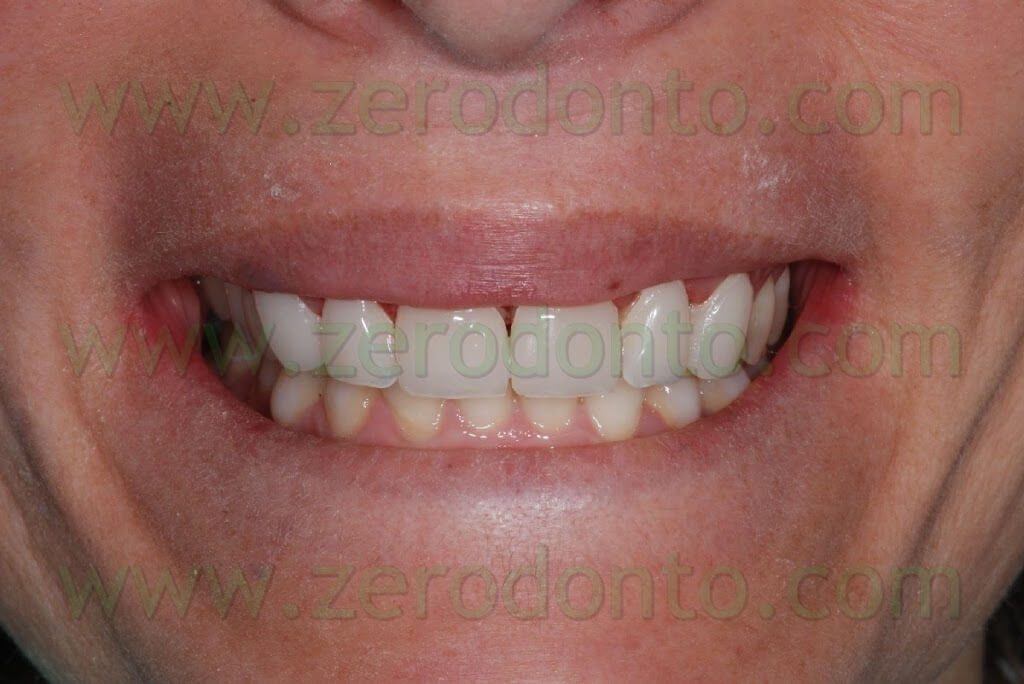
A female patient, aged 33, needed a functional and aesthetic improvement of the maxillary anterior region previously restored with composite veneers. The chair-side examination showed a marked discoloration of the restorations and an evident and generalized wear of the incisal edges of the anterior sectors of both the maxillary and mandibular jaws.
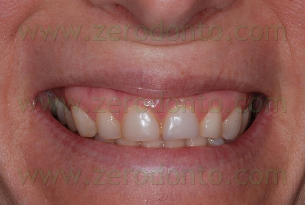
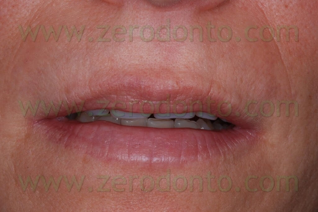
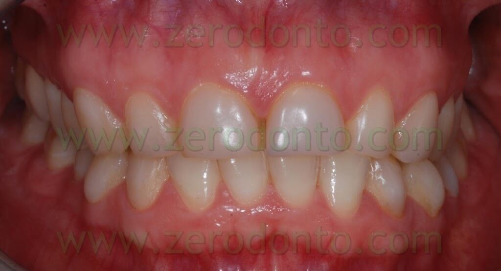
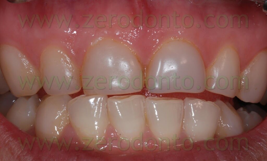
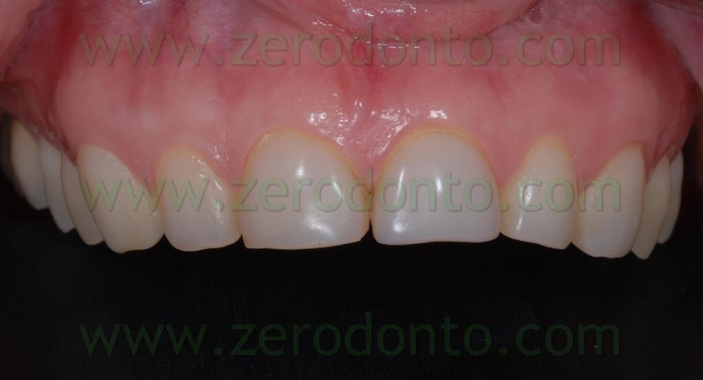
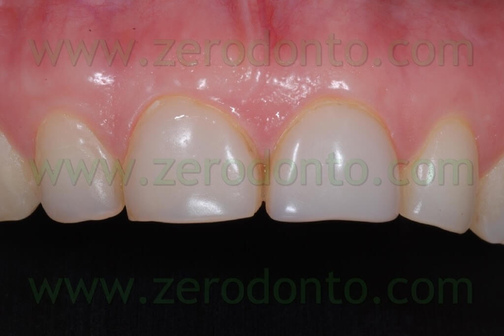
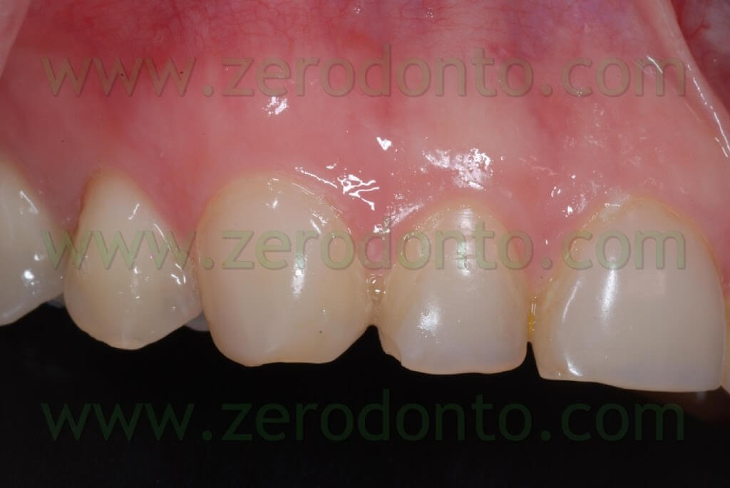
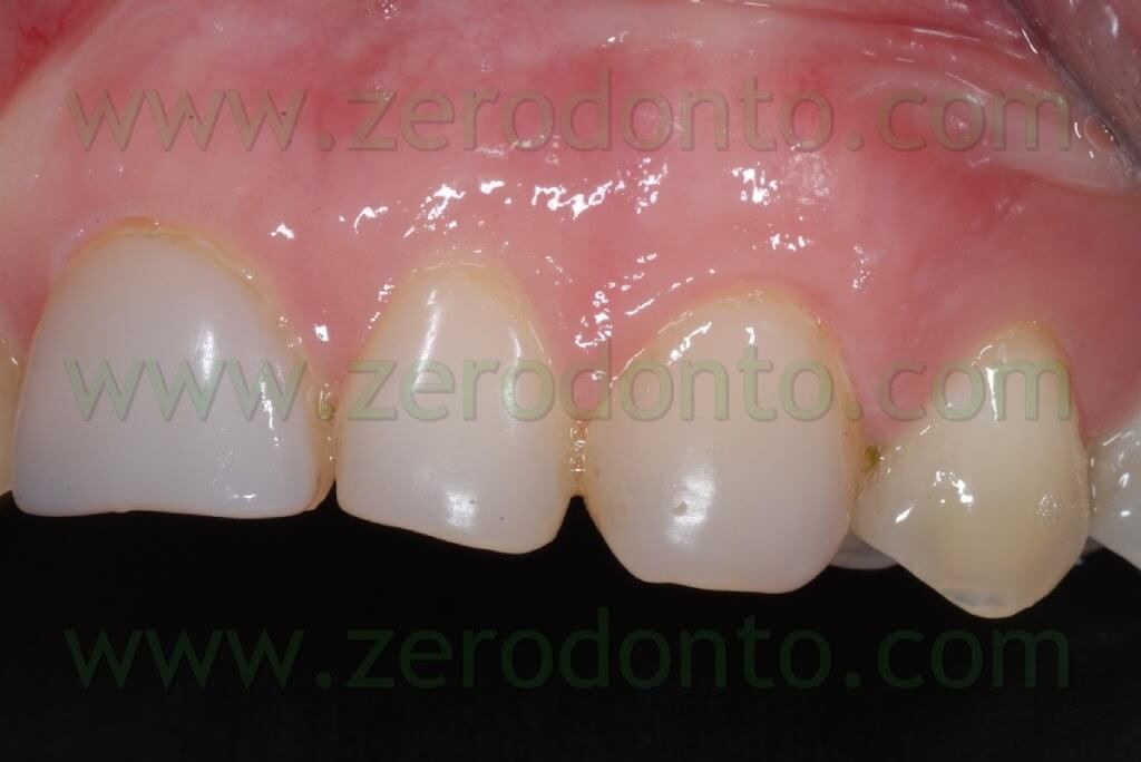
After explaining the different therapeutic options to the patient, it was decided to build-up 6 feldspatic porcelain veneers to restore elements 11, 12, 13, 21, 22 and 23. Moreover, it was planned to lengthen the incisal margins, in order to improve aesthetics, restore a correct protrusive function and optimize both the overjet and the overbite.
Evaluation of occlusal parameters is of primary importance for long term success of porcelain veneers. It is particularly important to assess the centric relation and protrusion in order to decide if and up to what point extending the palatal tooth preparation. In fact, the centric contacts have to be placed on porcelain, in order to avoid tensive stresses on the adhesive interface between the dental tissues and the restoration during protrusion.
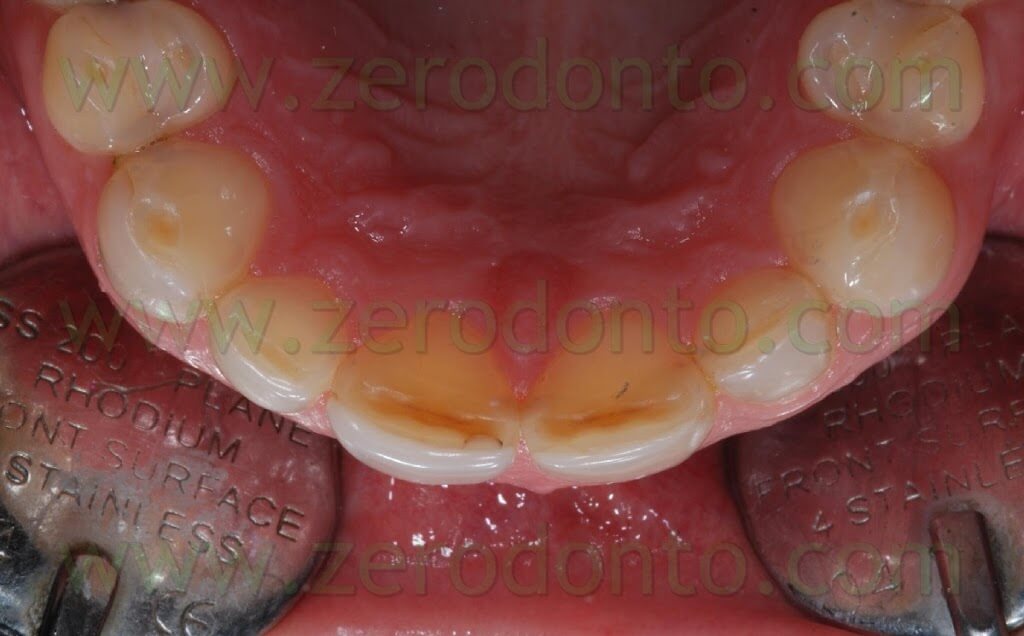
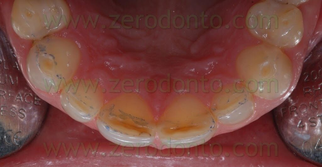
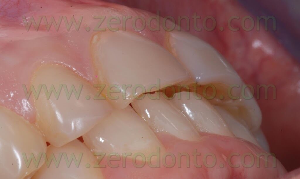
An alginate impression was taken in order to make a diagnostic model to allow the dental technician to make a mock up. The mock-up is a diagnostic aid useful to decide, together with the patient, shape, length and colour of the restorations.
Dental laboratory procedures
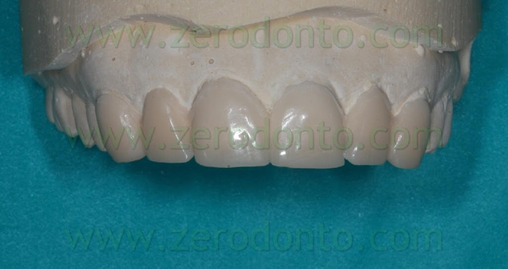
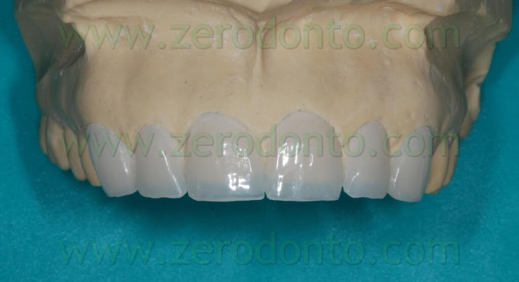
After receiving the alginate impression, some diagnostic models were made in the lab: the models were subsequently waxed-up to decide dimensions and volumes of the final restorations.
It is worth remembering that such parameters are dependent on the information received from the clinician. The dentist, in fact, will provide us with all the models alongside with centric, lateral and protrusive waxes – essential to make anterior guides – and specific pictures with detailed information on the anatomy and dynamics of facial and lip expressions and smile. All these data will let us make aesthetic and functional assessments useful to choose shape and to restore the length of the incisal margins – anatomical characteristics that, in this case, needed correction because of the presence of old and inadequate composite restorations.
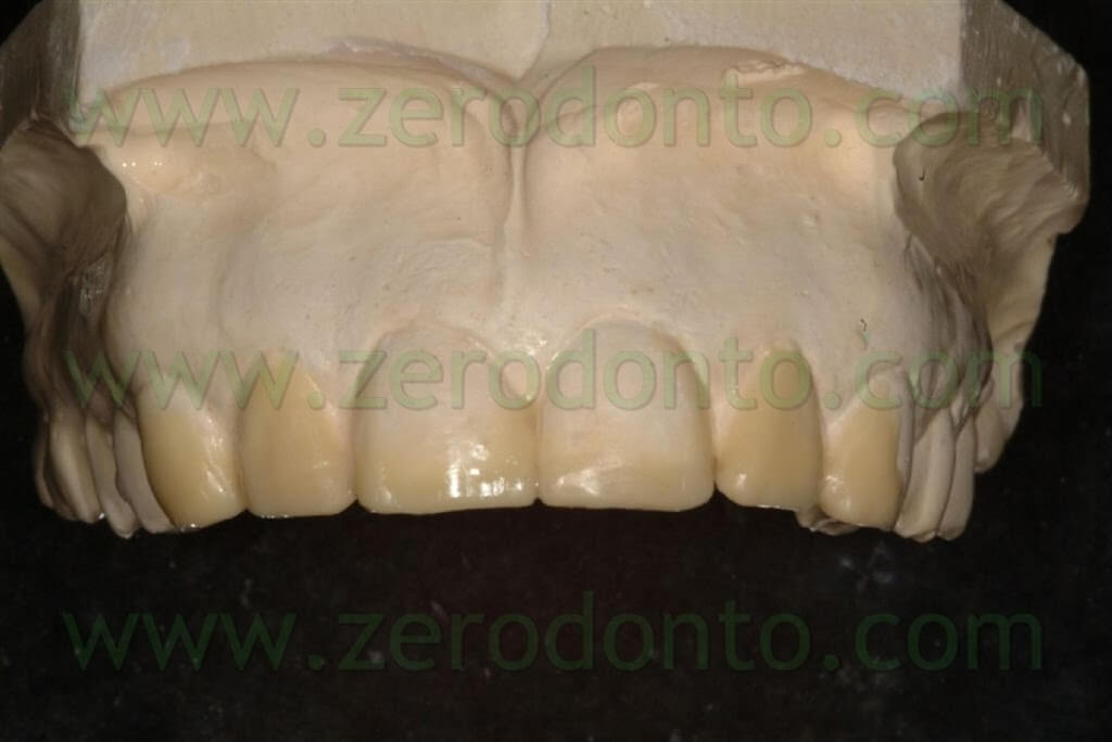
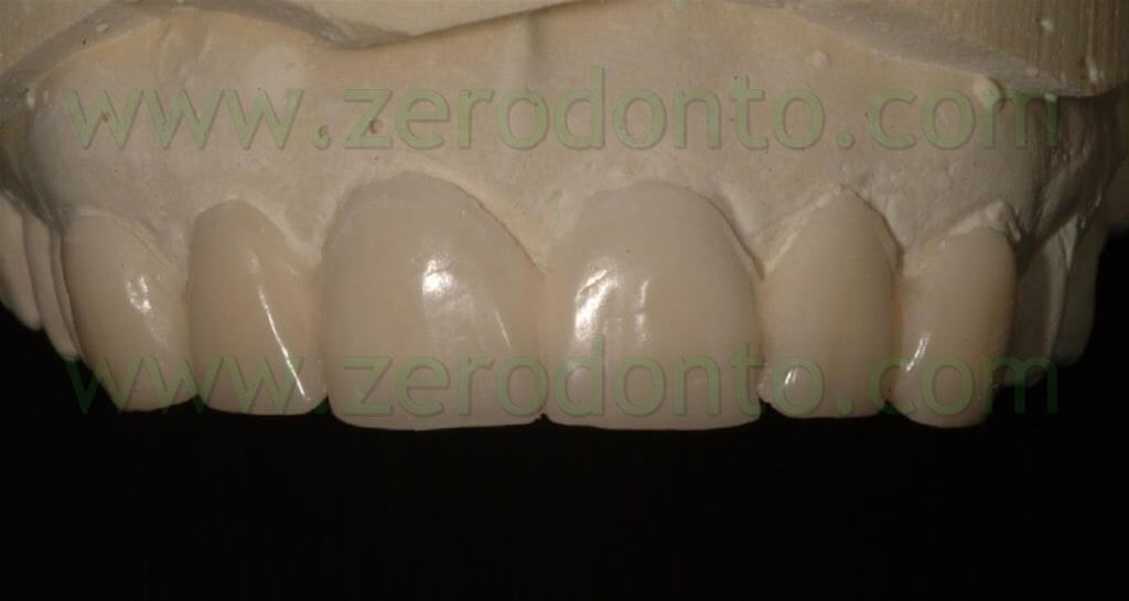
After completing the wax-up, we will go on to the subsequent making of the mock-up, test and control bench of our lab planning, that will provide us, while testing on the patient, all the information needed to make first the temporary and then the final restoration.
The great attention paid during the diagnostic phase is of primary importance to make our usual prosthetic restorations; it becomes even greater when the prosthetic option chosen is to make feldspatic veneers: their making, in fact, leaves no room for adjustments and improvised and offhand solutions that would impair the final result.
Clinical Part
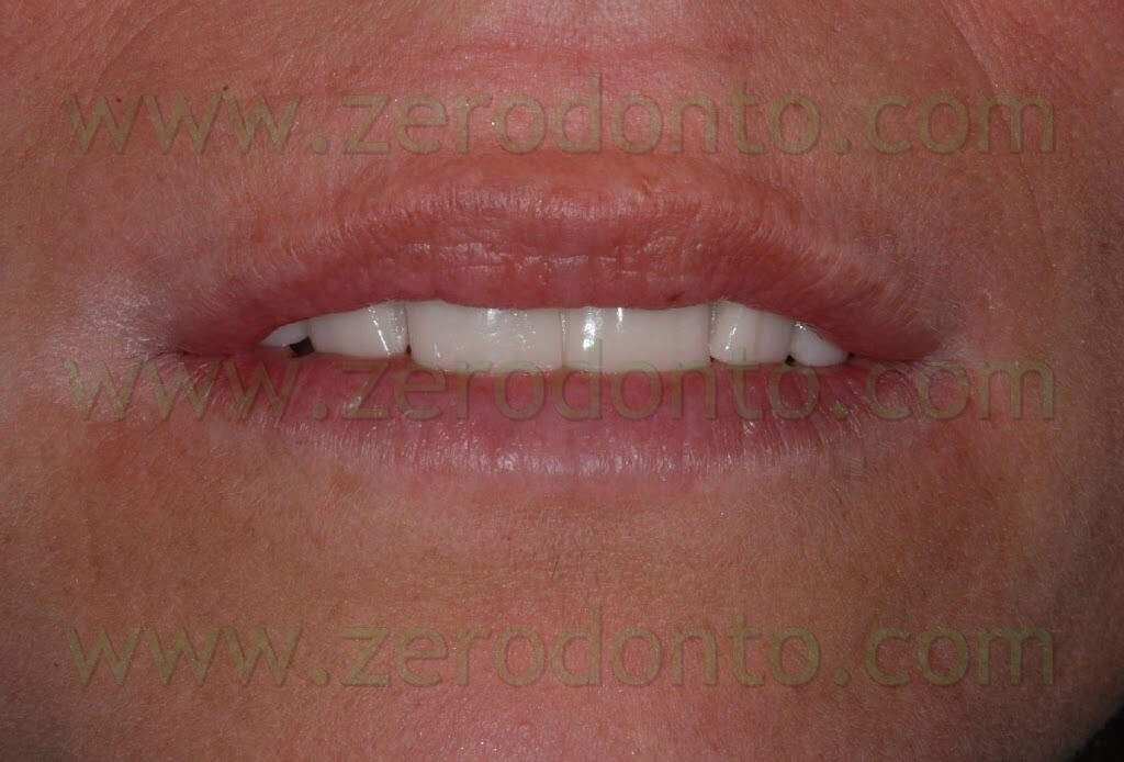
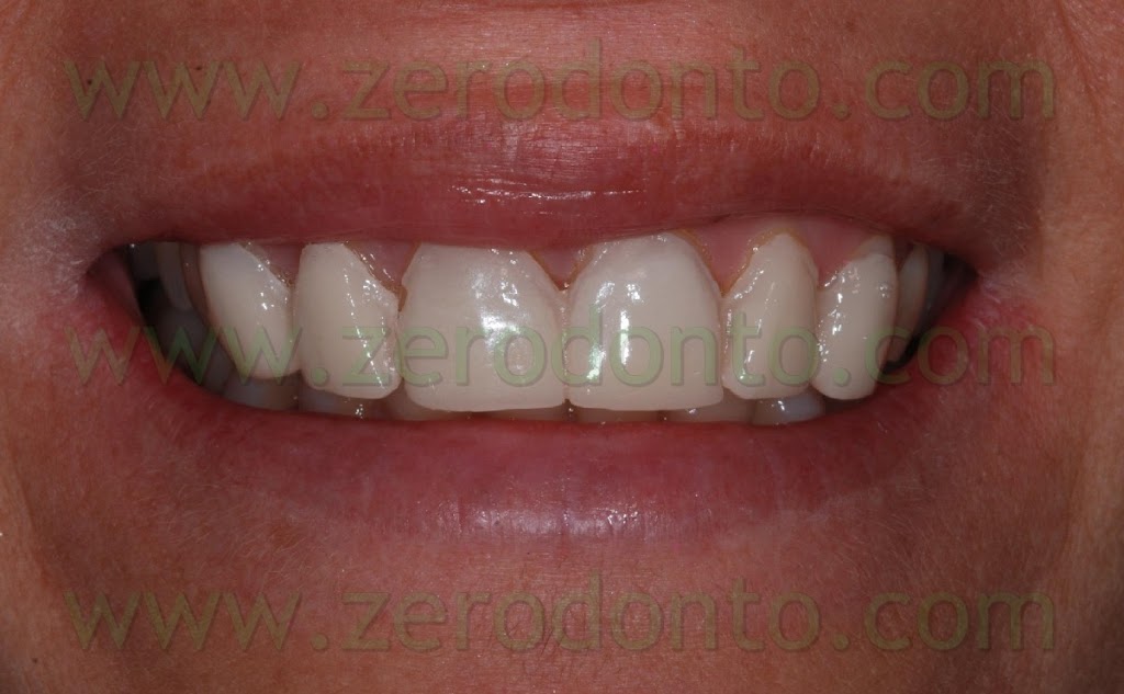
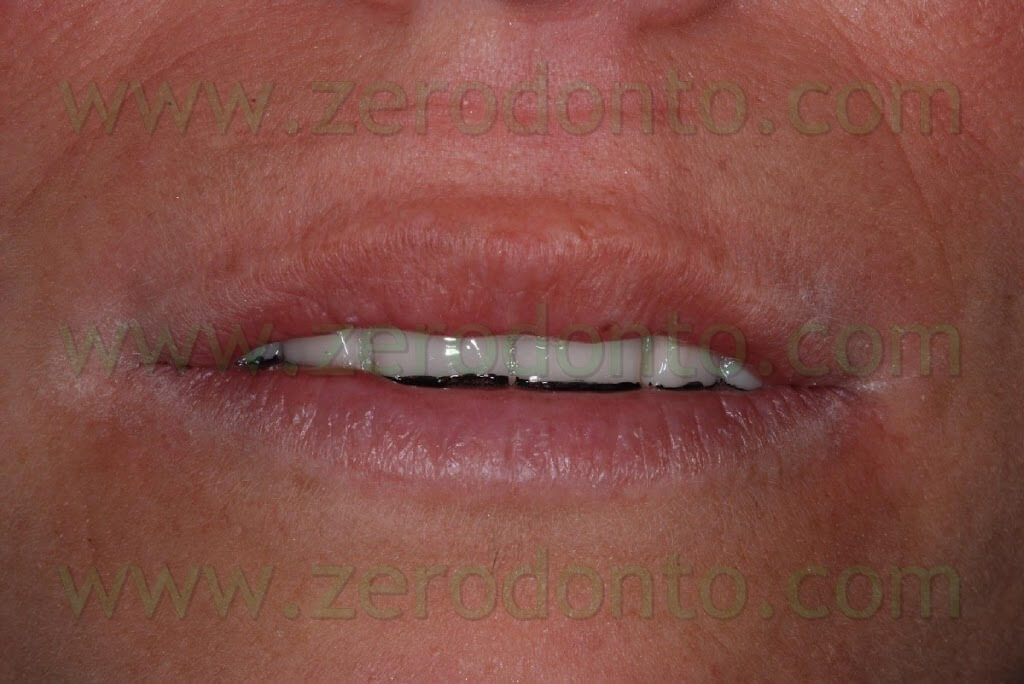
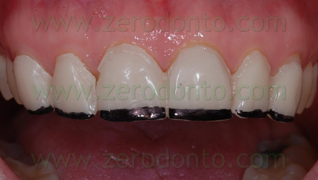
The mock-up is fit intraorally by simply applying it on the elements to be restored, without any fixing aid. Then the optimal length of the incisal margins is set and marked with black ink in order to have a clear visual contrast and to check the correct morphology of subsequent restorations. Once the correct length is set, it is possible to remove the part marked in black with rotary instruments in order to give the patient an idea of the forthcoming tooth morphology.
Dental laboratory procedures
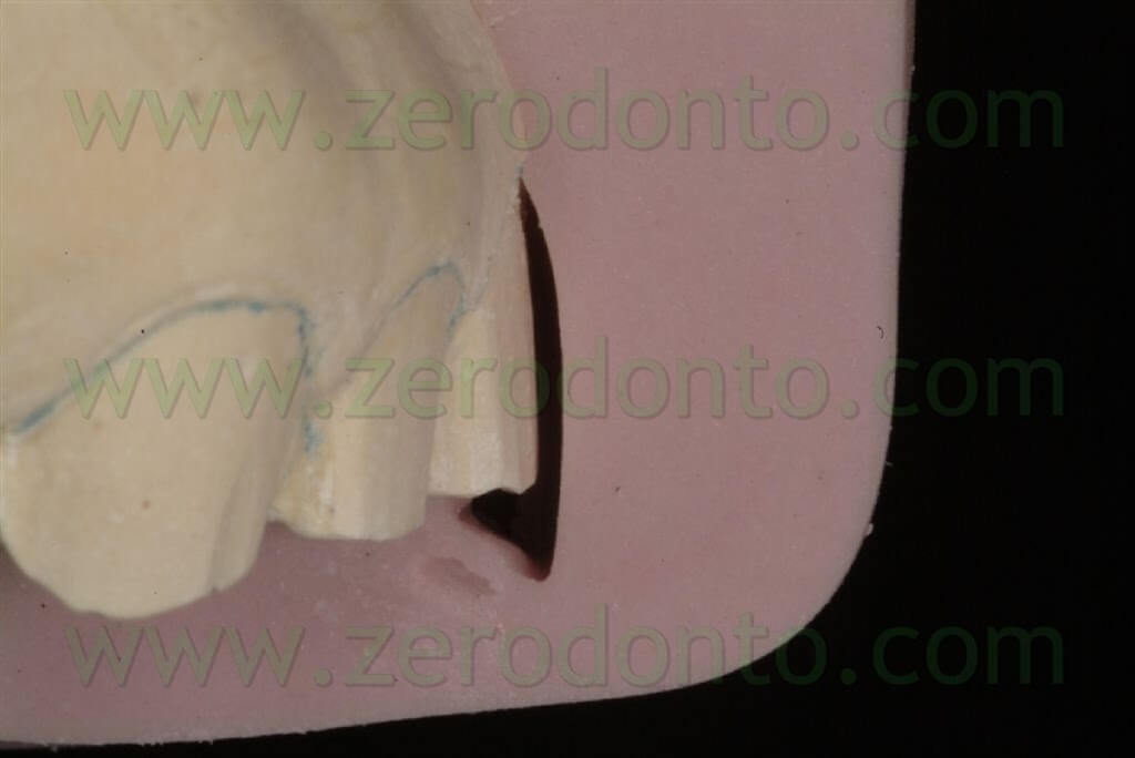
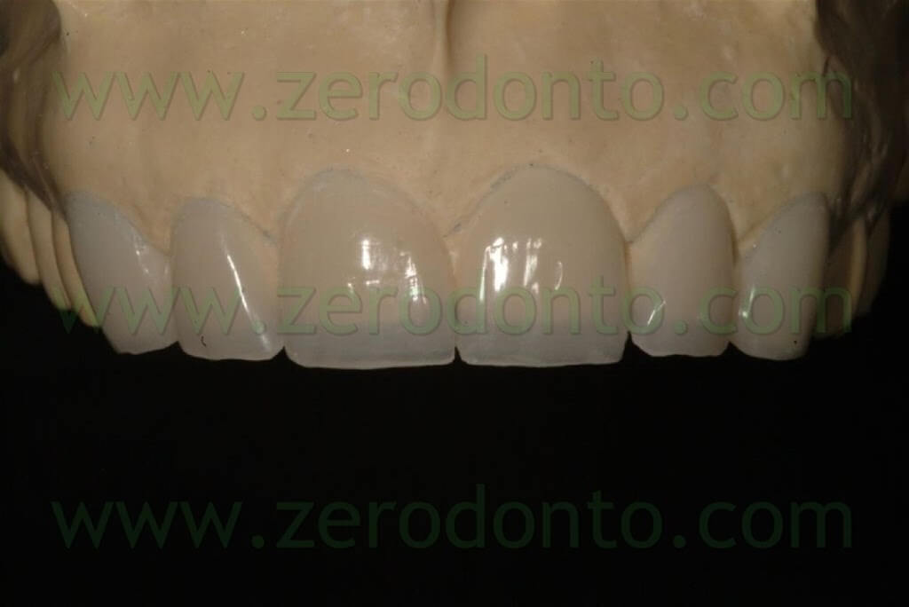
The temporary restoration will be made after trying the mock up: this trial can provide further information to make it. The temporary restoration, made of acrylic resin, will be built on a model upon which we will have already done some pre-filing adjustments: these are made using silicone guide masks obtained from the waxing and the mock-up, that may be modified, as in this case, after the trial.
Clinical part
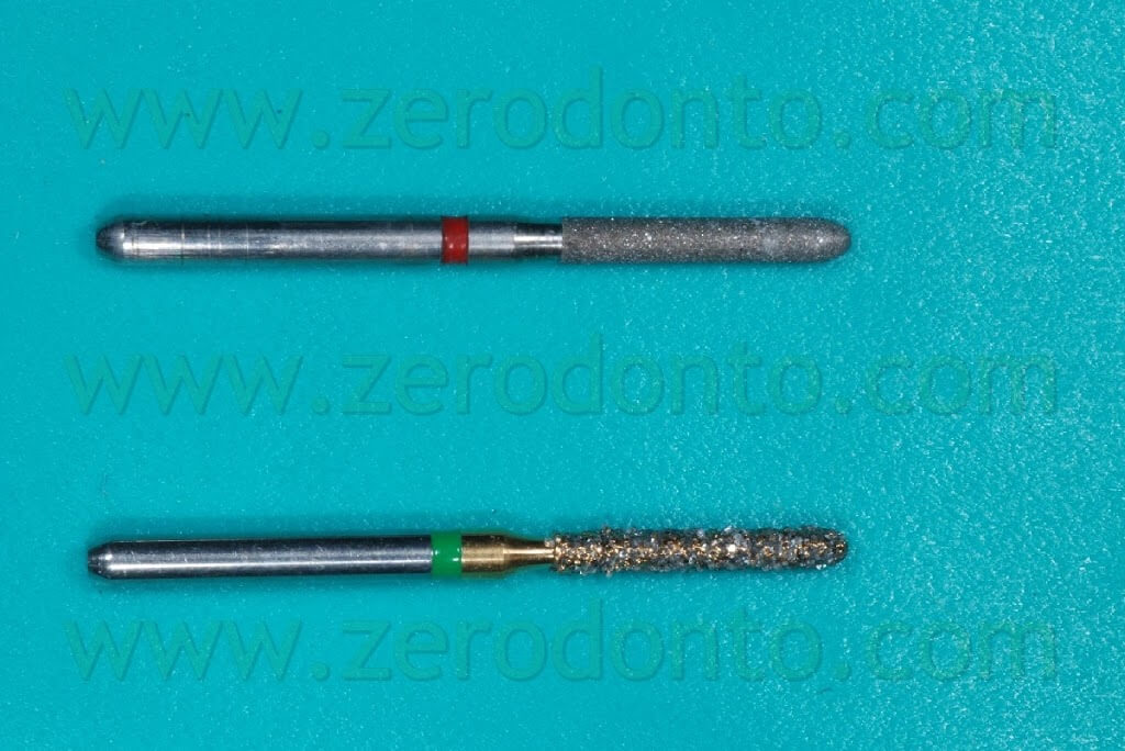
Before performing the dental preparations, we inserted intrasulcularly a multi-interweaved retraction chord for each dental element. Afterwards the preparations were made using a diamond, cylindrical dental drill with a round-edged, coarse-grained bur (0.14 of diameter) put up on a multiplier. A diamond, extra fine-grained dental drill with the same morphology was subsequently used to refine preparation geometries; it is possible to use, where necessary, an Arkansas stone to polish and regularize surfaces.
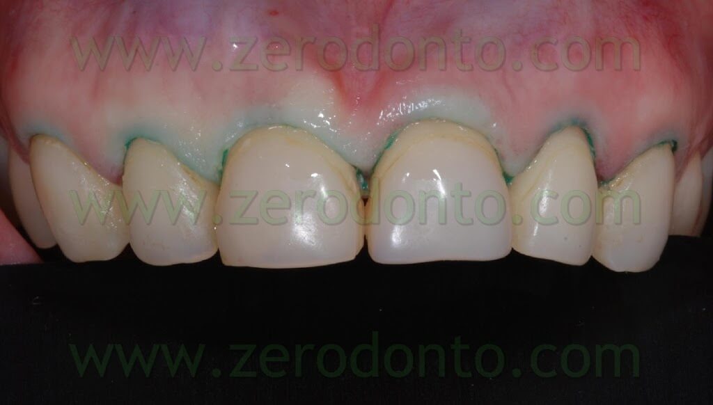
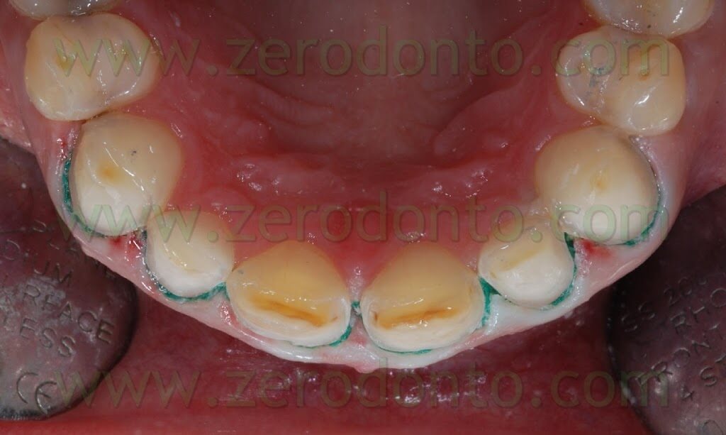
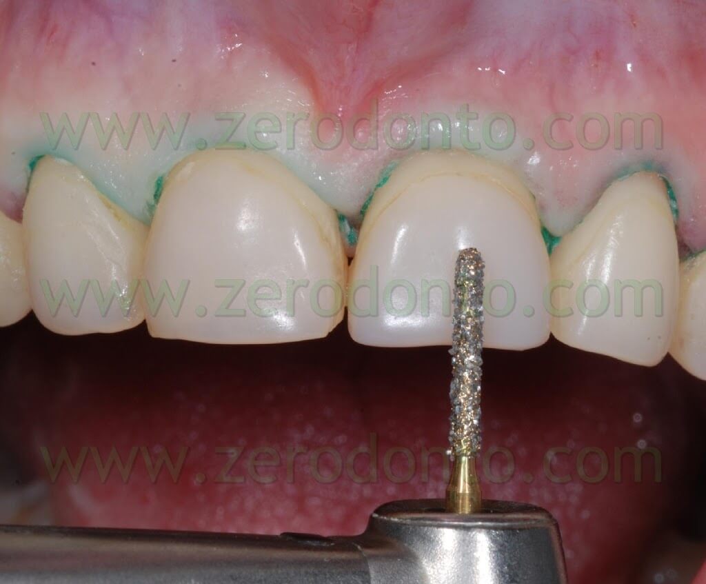
The preparations were made with a vestibular depth of about 0.7 mm, checked sinking the drill for half of its length during the preparation. We made palatal chamfer preparations extending for 2 mm coronally to the incisal margin on the palatal surface.
When clinical conditions allow it, this geometry of preparation may prove to be better than a window preparation limited to the buccal surface for a number of reasons:
- limited stress of the adhesive interface during protrusive function;
- possibility for the dental technician to deal at best with incisal transparencies of the restorations, without the necessity to mask residual dental tissue on the edge;
- univocal positioning of the restorations and reaching rock bottom during cementation.
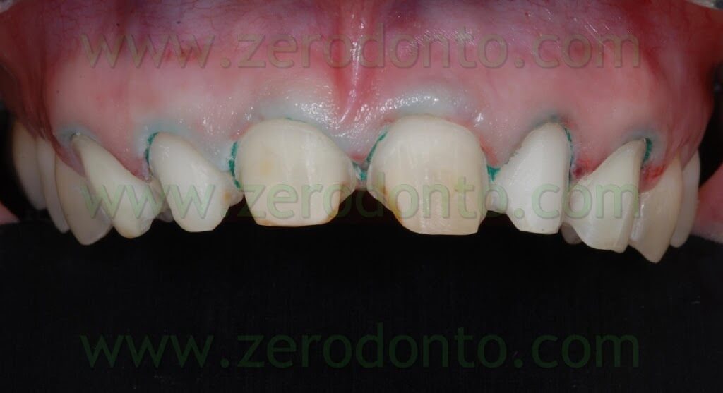
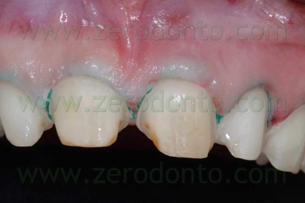

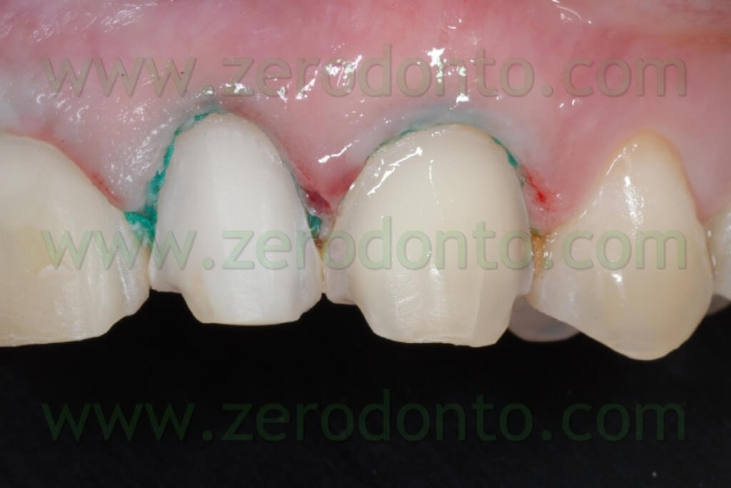
Previous composite restorations present on the elements to be restored were completely removed in the interproximal zones to ensure a correct prosthetic seal. On the contrary, on vestibular surfaces non infiltrated areas of composite were left in place in case their removal could have caused structural weakening of the dental elements. Present day literature agrees in stating that the presence of a composite surface equal or inferior to 30% of the whole adhesive surface does not represent a risk factor for the cementation of porcelain veneers.
The remaining composite can be either sandblasted intraorally or simply cleaned out and coarsened to help the subsequent bonding procedures.
Interproximal spaces were opened using a metallic abrasive strip on the sides of the enamel; this procedure helps in creating correct contact areas.
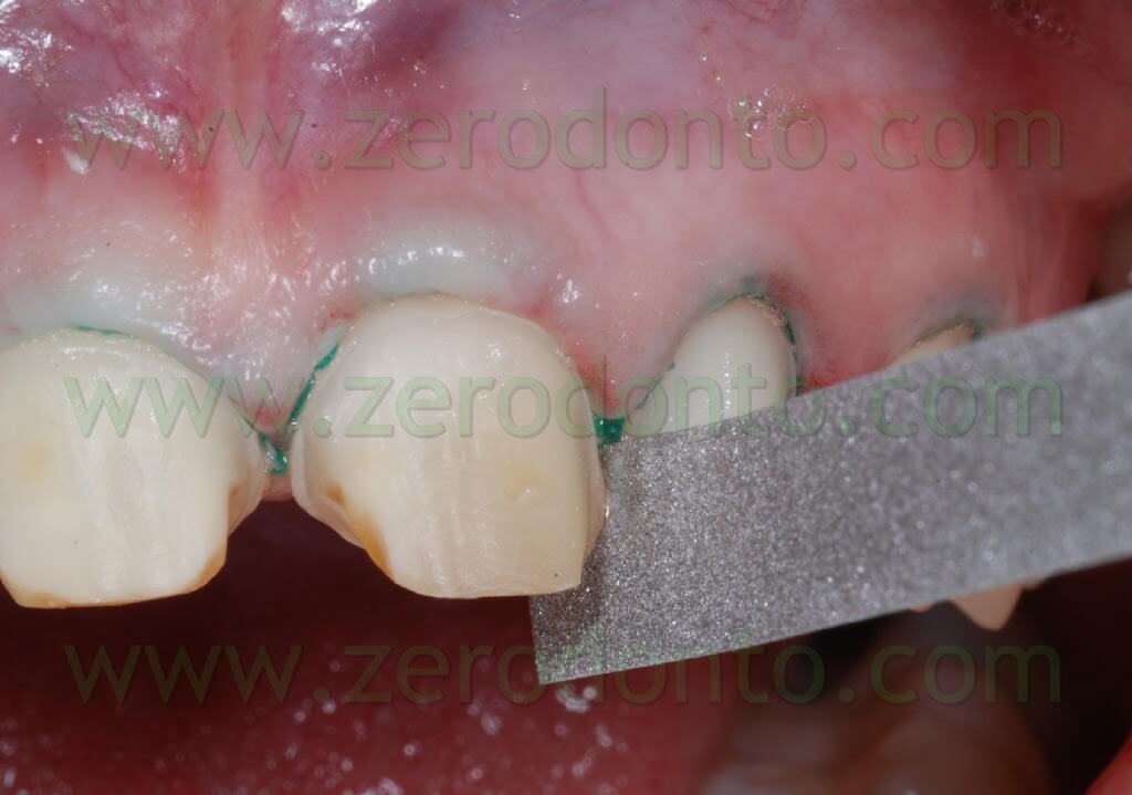
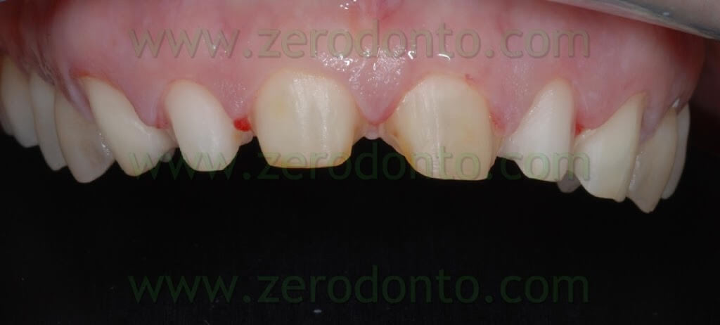
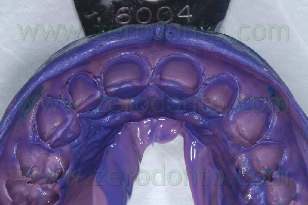
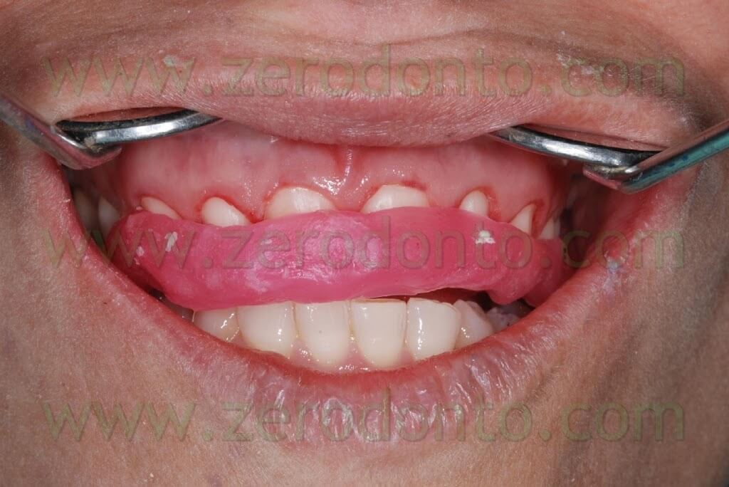
After finishing the dental preparations, a two-phase polyether impression was taken with a low-viscosity material injected intraorally on the preparations and a medium-viscosity material in the impression tray. Finally intermaxillary relations were recorded using centric wax on dental preparations.
Afterwards, the previously made temporary restoration was rebased intraorally with self-polymerizing acrylic resin. This temporary restoration was designed by joining all front elements in order to improve retention of the prosthesis, since porcelain veneers are restorations without primary retention. Once done, the temporary restoration was fixed intraorally with fluid composite, without etching the dental tissues.
Dental laboratory procedures
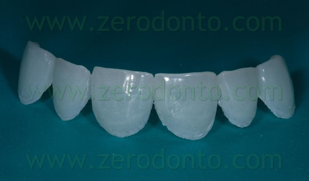
Making feldspatic porcelain veneers requires us to make several test models: master models in chalk and duplicate models in refractory material (coating). The coating used for refractory duplicates must have specific features needed for ceramic cooking.
- Excellent possibility to reproduce details
- Sufficient hardness during manipulation phases
- Possibility to dispose of polluting waste during degasification phase
- Expansion coefficient (C.T.E.) compatible with the one of the used ceramics
- Ease of removal of the veneers after their completion
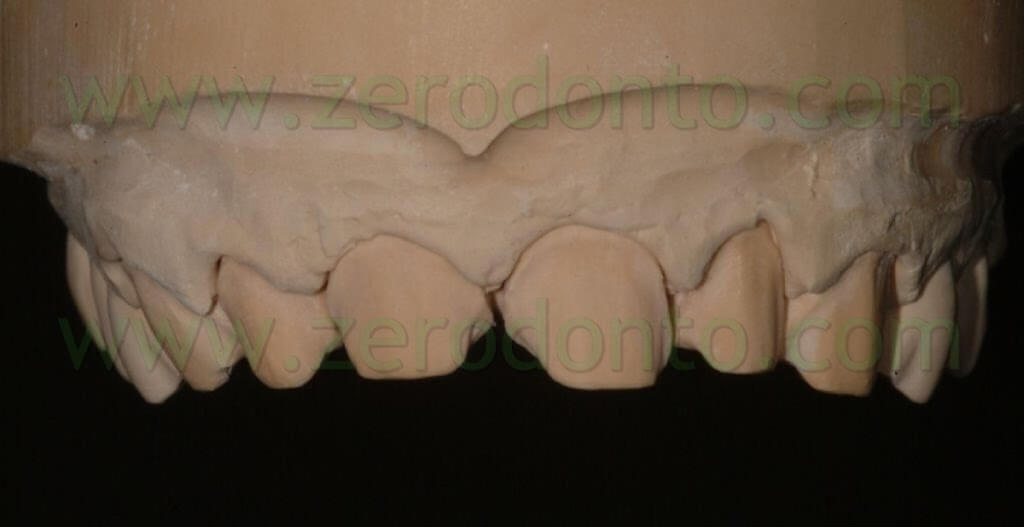
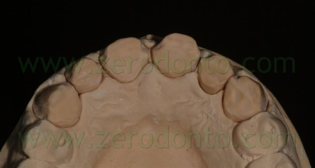
Once the model and the refractory duplicates are finished, first we will proceed to degasification, then to make the refractory models waterproof. This procedure consists in coating all the surface that duplicates the prepared zone with a layer of transparent ceramics.
This procedure has got a double function:
the first is to completely isolate the subsequent ceramic layers to be laid, protecting them against the absorbing action of the porous surface of the coating that would cause a constant loss of moisture during the phases of stratification;
the second is to create a peripheral contour zone of transparent ceramic mass that will hide more easily the transition zones between restoration and preparation (lens effect).
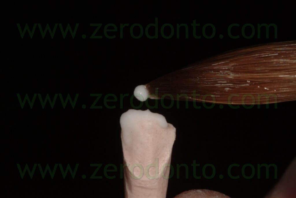
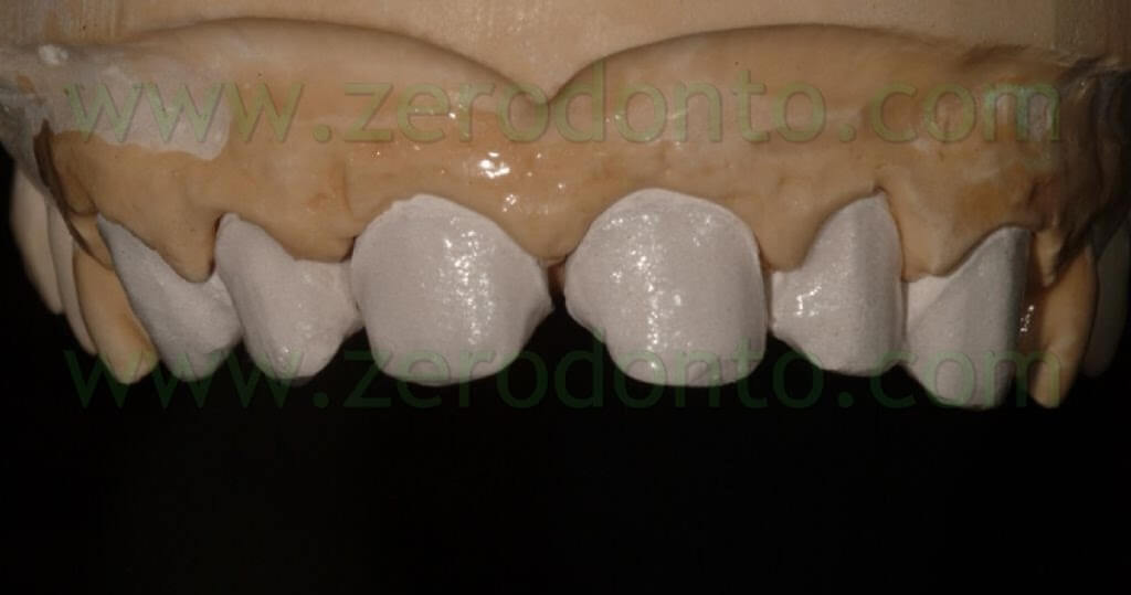
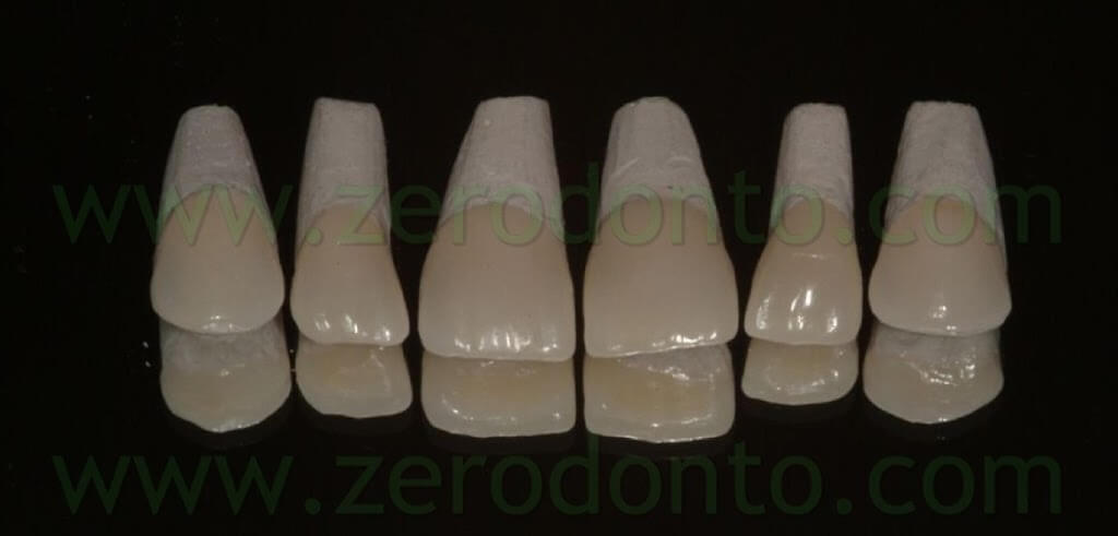
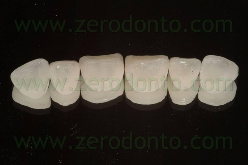
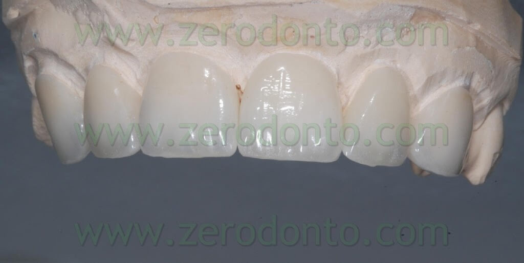
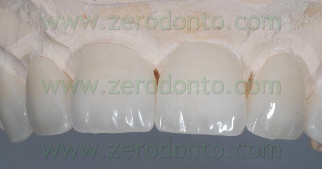
We will finally go on to simplified stratification, at the end of which, after verifying intraoral aesthetics, it will be possible to free ceramics from the refractory models, to check contact points on master models and to send them to the dental office for cementation.
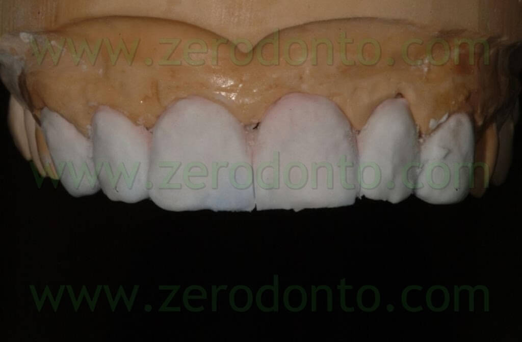
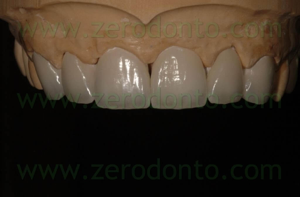
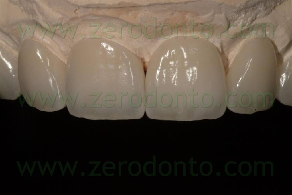
Clinical part
7 days after putting the temporary restoration in place, we checked colour and aesthetics of the veneers. They were made of feldspatic ceramics, since it provides excellent aesthetics and can be etched, while all-ceramic alumina and zirconia based ceramics cannot be chemically etched. Use of an etching agent on feldspatic ceramics determines the creation of superficial cavitations which increase micromechanical retention of the restorations. Before getting to the dental office, the restorations were sandblasted in the lab.

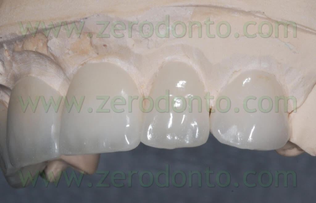
The following step was cementation using a dedicated resinous dual cement.
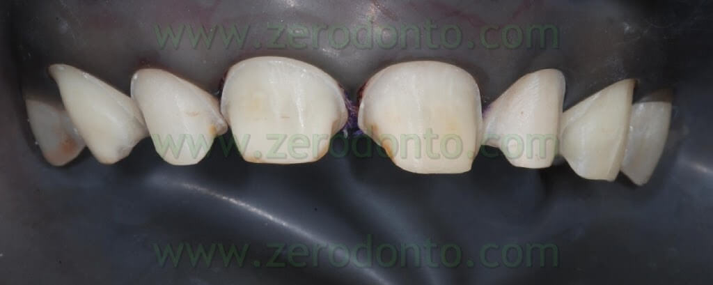
The operative area was isolated with a rubber dam without clamps: the edges of the dam sheet were invaginated intrasulcularly using a multi-interweaved retraction chord around every dental element.
This chord is also useful to help removal of excess cement.
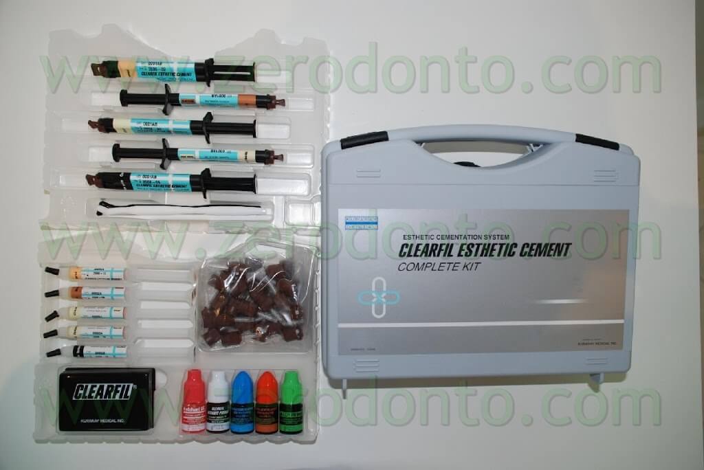
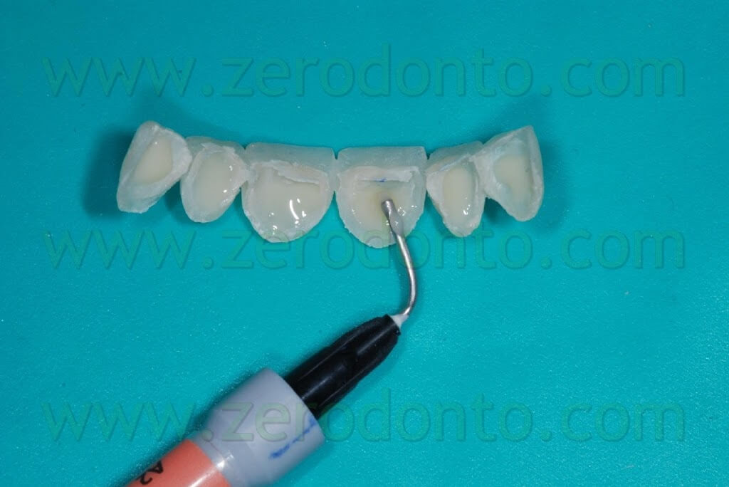
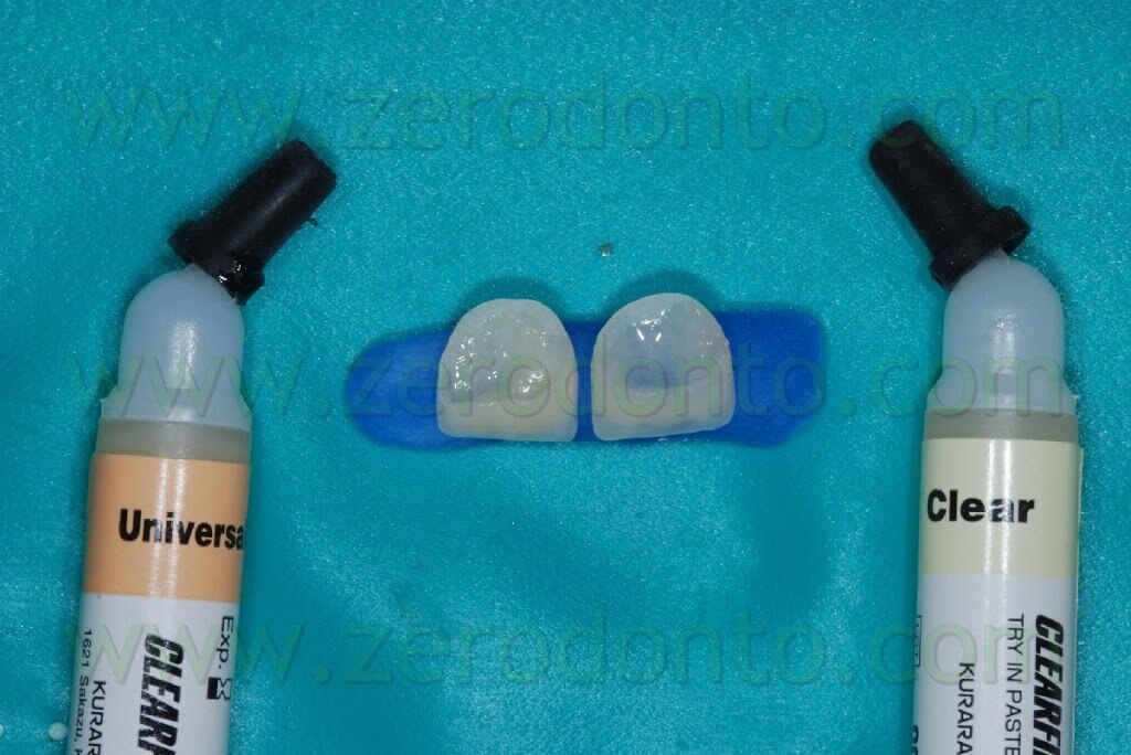
Cement colour was chosen using two water-soluble test pastes put inside the veneers covering elements 11 and 21: on element 11 we used a “clear” (transparent) tone, while on element 21 we used a “universal” tone (universal). In agreement with the patient, we chose to use the “universal” tone for cementation.
Thanks to its being completely water-soluble, it is possible to dispose eventual paste waste with a simple rinse and drying.
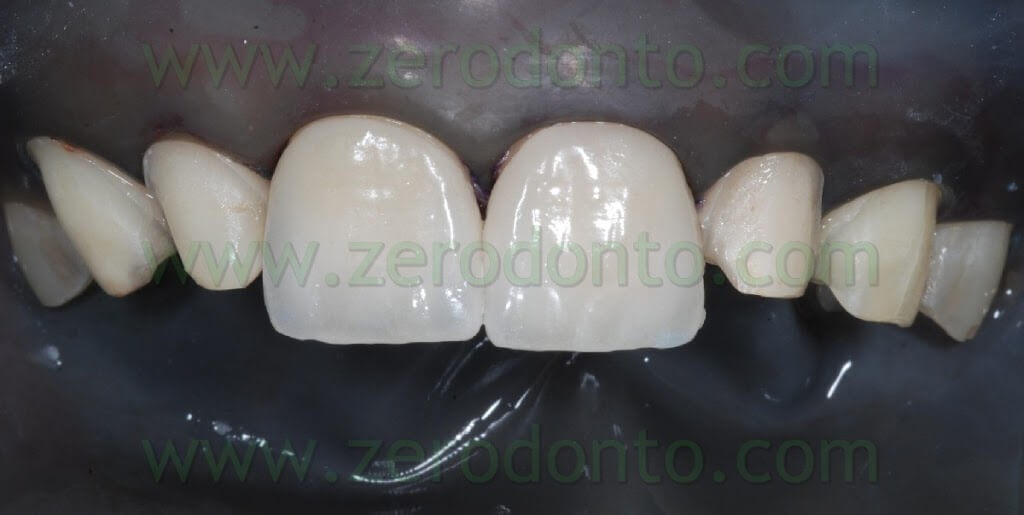
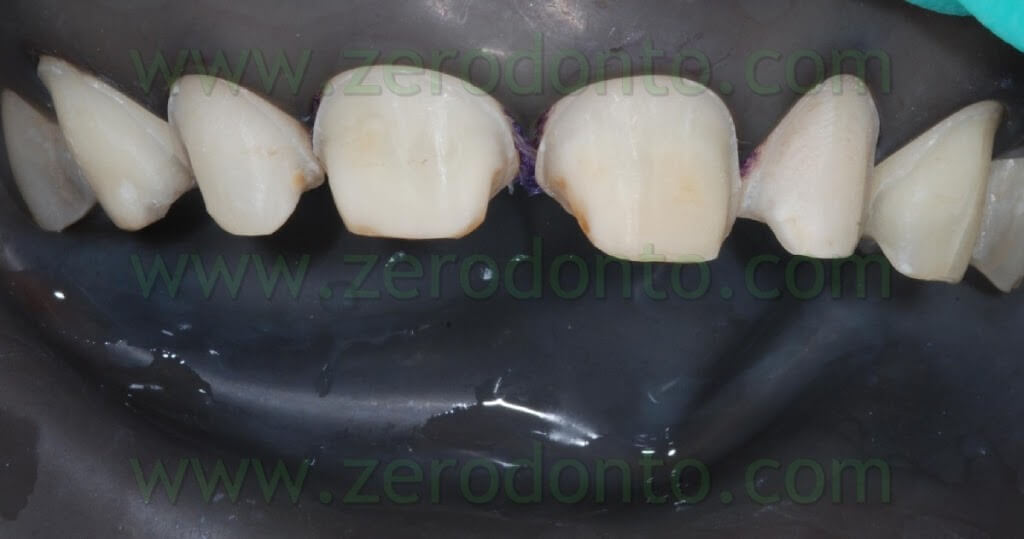
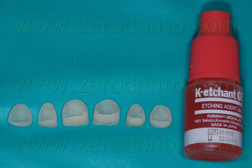
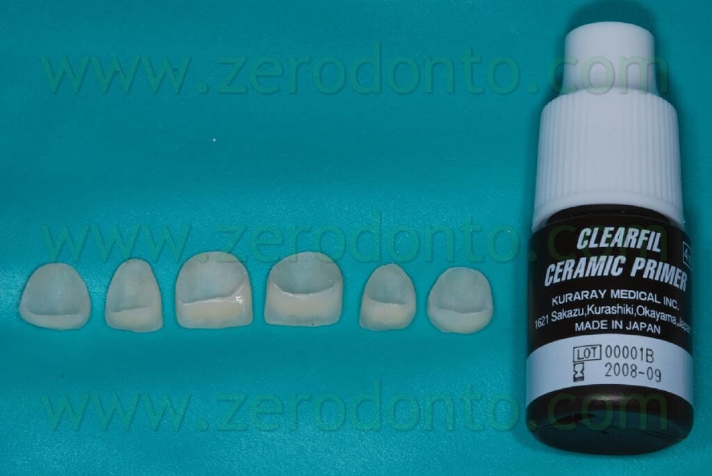
Following the protocol, we used an etching agent for 10 seconds on dental elements and 5 seconds on the internal surfaces of the veneers. Once rinsed and dried, internal surfaces of the restorations were conditioned with a ceramic primer.
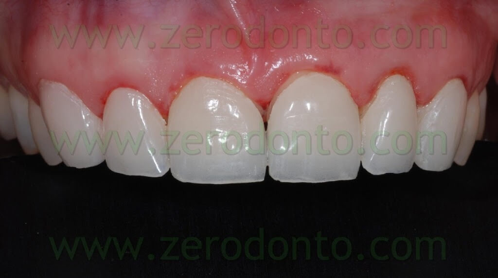
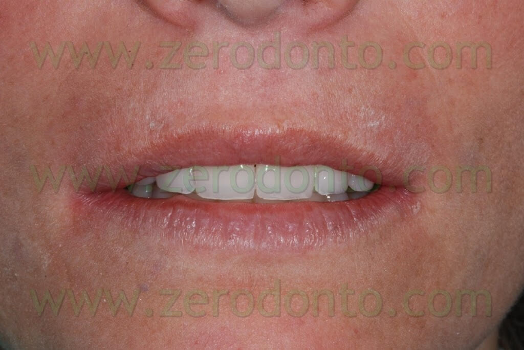
Finally, the six veneers were cemented and the usual operations of removal of excess cement and, where necessary, refining and polishing of the edges using small rubber wheels for ceramics were carried out.
Particular care was dedicated to centric, protrusive and lateral occlusive control.
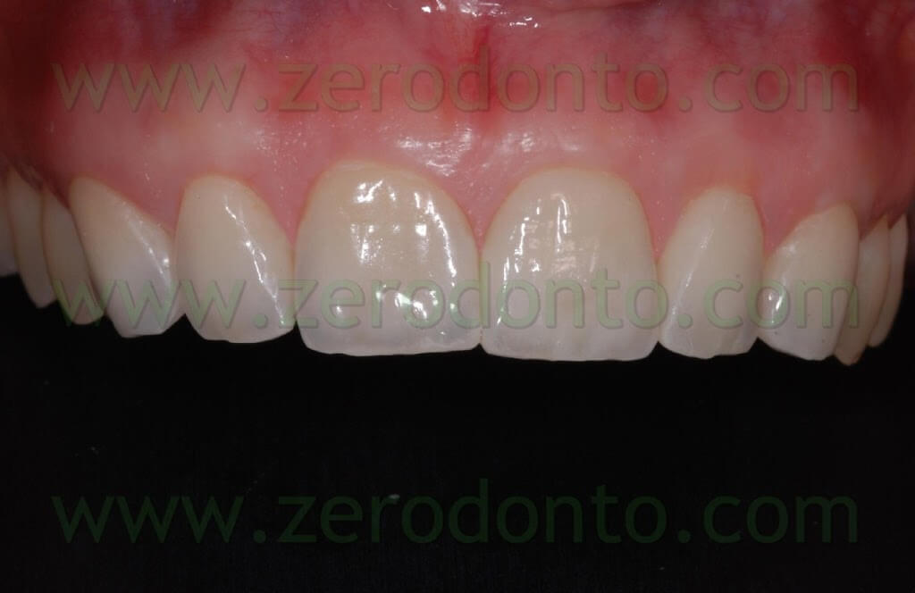
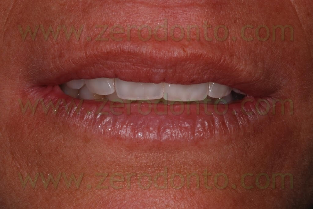
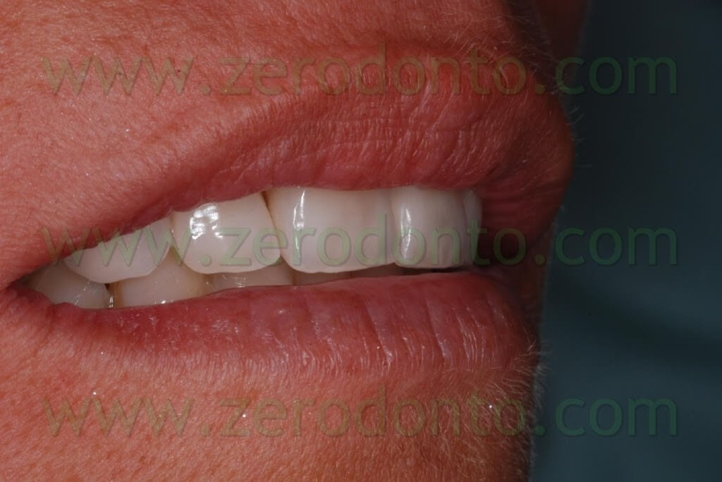
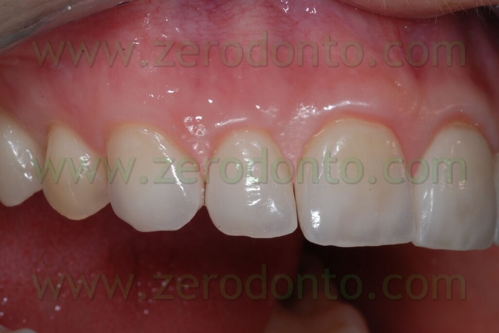
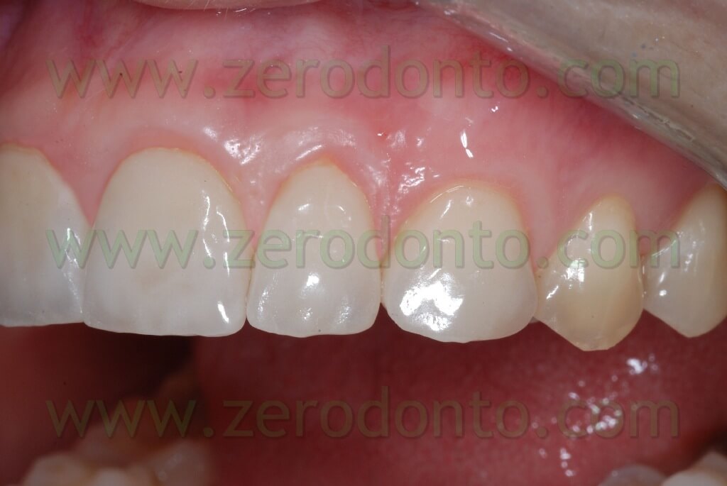
BEFORE

AFTER

BEFORE

AFTER

It is advisable not to use toothpastes containing fluorine or fluorinated agents for the 2 weeks prior to the cementation, in order to avoid a hyper-mineralization of the dental tissue that may interfere with the sticking.
Bibliography
- Sadowsky SJ.
An overview of treatment considerations for esthetic restorations: a review of the literature.
J Prosthet Dent. 2006 Dec;96(6):433-42. Review.
- Magne P.
Immediate dentin sealing: a fundamental procedure for indirect bonded restorations.
J Esthet Restor Dent. 2005;17(3):144-54; discussion 155. Review.
- Wakiaga J, Brunton P, Silikas N, Glenny AM.
Direct versus indirect veneer restorations for intrinsic dental stains.
Cochrane Database Syst Rev. 2004;(1):CD004347. Review.
- Strassler HE.
Minimally invasive porcelain veneers: indications for a conservative esthetic dentistry treatment modality.
Gen Dent. 2007 Nov;55(7):686-94; quiz 695-6, 712.
- Smales RJ, Etemadi S.
Long-term survival of porcelain laminate veneers using two preparation designs: a retrospective study.
Int J Prosthodont. 2004 May-Jun;17(3):323-6.
- Zarone F, Epifania E, Leone G, Sorrentino R, Ferrari M.
Dynamometric assessment of the mechanical resistance of porcelain veneers related to tooth preparation: a comparison between two techniques.
J Prosthet Dent. 2006 May;95(5):354-63.
- Peumans M, De Munck J, Fieuws S, Lambrechts P, Vanherle G, Van Meerbeek B.
A prospective ten-year clinical trial of porcelain veneers.
J Adhes Dent. 2004 Spring;6(1):65-76.
- Magne P, Belser UC.
Novel porcelain laminate preparation approach driven by a diagnostic mock-up.
J Esthet Restor Dent. 2004;16(1):7-16; discussion 17-8. Review.
- Zarone F, Apicella D, Sorrentino R, Ferro V, Aversa R, Apicella A.
Influence of tooth preparation design on the stress distribution in maxillary central incisors restored by means of alumina porcelain veneers: a 3D-finite element analysis.
Dent Mater. 2005 Dec;21(12):1178-88. Epub 2005 Aug 10.
- Magne P, Perroud R, Hodges JS, Belser UC.
Clinical performance of novel-design porcelain veneers for the recovery of coronal volume and length.
Int J Periodontics Restorative Dent. 2000 Oct;20(5):440-57.
- Mutone V.
L’integrazione Bioestetica
Dental Labor 2005 Mag.
- Mutone V.
Stratificazione semplificata.
Rivista di tecnologie dentali Feb. 2003
- Piconi C., Rimondini L., Cerroni L., Donati C., Mutone V.
La Zirconia in Odontoiatria
- Mauro Fradeani
La riabilitazione estetica in protesi fissa- Vol. I
Analisi estetica
For information:
Mr. Vincenzo Mutone
Mr. Mutone was born on January 20th, 1965 and obtained his qualification as a dental technician in Naples IPSIA “Casanova”.
He holds a lab since 1983.
He took part in many courses in Italy and abroad among which some with Klaus Muetherties and Willi Geller, from whom he learnt his practical teaching and aesthetic philosophy attending many times his lab in Zurich (CH). He was business partner and co-owner of the Oral-Design 2 lab along with Mr. Giuseppe Zuppardi in the years 1994-96. When this experience was over he met professionally Mr. Atoshi Aoshima, who led him to appreciate Japanese aesthetic school. After this experience he started another project that led him to make a systematics for ceramic masses multistratification for the Noritake Kizai, LTD company (Japan).
In the last decade he has held conferences and communications on metal ceramics and aesthetics in many national and international meetings.
Today he focuses particularly on prosthesis implant and aesthetics using modern materials such as zirconia and CAD-CAM methods. He is also taking part in projects that aim to carry out and spread implantology based on computer planning with immediate function application and in the making of a multistratification system on zirconia oxide structures.
For information:
zerodonto@gmail.com
Website: www.vincenzomutone.it
vincenzomutone@virgilio.it

